retuve.docs.overviews.clinicians
Retuve for Clinicians
Retuve is made up of two main analysis modes:
- DDH Ultrasound: the Standard Alpha Angle and Coverage, as well as other more complicated 3D measurements.
- DDH X-Ray: Currently just acetabular index.
DDH Ultrasound
Retuve runs 3 seperate DDH Ultrasound Models:
References to more specifics on these indicies can be found here: https://boneandjoint.org.uk/Article/10.1302/2633-1462.311.BJO-2022-0081.R1/pdf
3D Ultrasound
Please Note that the 2D Sweep and 3D DICOMs are sythetically stitched together from 2D Graf Hips, so do not expect accurate results. We are working on getting a 3DUS Case on a Creative Commons License to have as an example.
Retuve makes the following measurements in a 3D Hip Ultrasound:
- Alpha Angle
- Coverage
- Curvature (A measure of the shape of the acetabulum/illium bone)
- ACA (Acetabular Coverage Angle) (A more precise 3D Alpha Angle)
- Centering Ratio (A measure of the femoral head position in 3D space)
We calculate the 2D measurements in 3D by finding the average in the:
- Graf Plane (including +/- 5% in average)
- Posterior
- Anterior
Alpha, Coverage and Curvature are visible on videos created by retuve, which can be seen here: (If you are on Github: see https://github.com/radoss-org/radoss-creative-commons/raw/main/other/ultrasound/172535.mp4)
Attribution of the above Ultrasound Images: Case courtesy of Ryan Thibodeau from https://radiopaedia.org 172535 (https://radiopaedia.org/cases/172535)
Attribution of the above Ultrasound Images: Case courtesy of Ryan Thibodeau from https://radiopaedia.org 172535 (https://radiopaedia.org/cases/172535)
The Centering Ratio and ACA values are not as easy to visualise, but can be seen in the following 3D Figure: (If you are on Github: see https://html-preview.github.io/?url=https://github.com/radoss-org/radoss-creative-commons/raw/main/other/ultrasound/172535.html)
Derivative from Radiopedia: Case courtesy of Ryan Thibodeau from https://radiopaedia.org 172535 (https://radiopaedia.org/cases/172535)
2D Ultrasound
Retuve makes the following measurements in a 2D Hip Ultrasound:
- Alpha Angle
- Coverage
- Curvature (A measure of the shape of the acetabulum/illium bone)
They can all be seen on the below image.
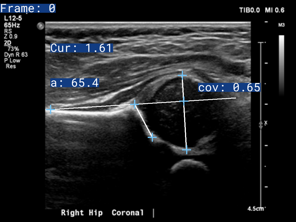
Attribution of the above Ultrasound Images: Case courtesy of Ryan Thibodeau from https://radiopaedia.org 172535 (https://radiopaedia.org/cases/172535)
2D Sweep Ultrasound (Experimental/Limited Testing)
Please Note that the 2D Sweep and 3D DICOMs are sythetically stitched together from 2D Graf Hips, so do not expect accurate results. We are working on getting a 3DUS Case on a Creative Commons License to have as an example.
2D Sweep Ultrasound is essentially 2D Ultrasound, with the added ability to detect which frame of a video/dicom to select for analysis. This is useful for scans that have not been processed already.
DDH X-Ray
Retuve runs a single DDH X-Ray Model, which measures the Acetabular Index.
This is visualised by an image created when Retuve Runs.

Attribution of the above X-Ray Images: Fraiwan, Mohammad; Al-Kofahi, Noran; Hanatleh, Omar; ibnian, ali (2022), “A dataset of DDH x-ray images”, Mendeley Data, V2, doi: 10.17632/jf3pv98m9g.2
 1# Copyright 2024 Adam McArthur
 2#
 3# Licensed under the Apache License, Version 2.0 (the "License");
 4# you may not use this file except in compliance with the License.
 5# You may obtain a copy of the License at
 6#
 7#     http://www.apache.org/licenses/LICENSE-2.0
 8#
 9# Unless required by applicable law or agreed to in writing, software
10# distributed under the License is distributed on an "AS IS" BASIS,
11# WITHOUT WARRANTIES OR CONDITIONS OF ANY KIND, either express or implied.
12# See the License for the specific language governing permissions and
13# limitations under the License.
14
15"""
16.. include:: ../../../docs/overviews/for_clinicians.md
17"""